 etcconseil Latest Updated Live News From etcconseil
Diagnostic ultrasound is a powerful medical imaging technique that is widely used to visualize internal structures of the body. It is commonly used in the field of radiology to diagnose and monitor various conditions. However, like any imaging modality, diagnostic ultrasound is not without its limitations and challenges. One such challenge is the occurrence of noise artifacts, which can affect the quality and accuracy of the ultrasound images.
Table of Contents
Noise Artifacts in Diagnostic Ultrasound
Noise artifacts are unwanted patterns or distortions that can appear on an ultrasound image, interfering with the clarity and diagnostic value of the scan. These artifacts can arise from various sources, including electronic interference, patient motion, and ultrasound system settings. They can manifest as speckles, lines, or other irregular patterns on the image.
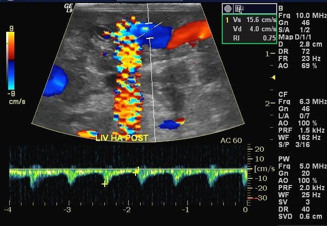
One common type of noise artifact is speckle noise, which appears as a grainy or mottled texture on the ultrasound image. Speckle noise can be caused by the interference of sound waves reflected back from the tissues being imaged. This interference leads to constructive and destructive interference patterns, resulting in the speckle pattern seen on the image.
Another type of noise artifact is motion artifacts, which occur when there is movement of the patient or the ultrasound probe during the scan. Patient motion can cause blurring or ghosting of structures on the image, making it difficult for the radiologist to interpret the findings accurately. Motion artifacts are particularly common in pediatric patients or individuals who are unable to remain still during the imaging procedure.
To minimize noise artifacts, radiologists and sonographers employ various techniques and strategies. Optimizing the ultrasound system settings, such as adjusting the gain, time-gain compensation, and focal zones, can help reduce artifacts. Additionally, ensuring proper patient positioning and minimizing patient motion can significantly improve image quality and reduce motion artifacts.
The Role of Magnetic Resonance Imaging
While diagnostic ultrasound is a valuable imaging modality, there are situations where magnetic resonance imaging (MRI) plays a crucial role in the diagnosis and management of certain conditions. MRI uses strong magnetic fields and radio waves to generate detailed images of the body’s internal structures. Unlike ultrasound, MRI does not use ionizing radiation and can provide multiplanar imaging with excellent soft tissue contrast.
Magnetic resonance imaging is particularly useful in the evaluation of neurological disorders, such as brain tumors, multiple sclerosis, and spinal cord injuries. It can also provide valuable information about musculoskeletal conditions, cardiovascular diseases, and abdominal and pelvic pathologies.
Furthermore, MRI can help identify and characterize various abnormalities, such as tumors, inflammation, and vascular malformations. It is often used as a problem-solving tool when other imaging modalities, such as ultrasound or computed tomography (CT), are inconclusive or insufficient.
In conclusion, diagnostic ultrasound is a valuable imaging tool that can provide real-time visualization of internal structures. However, the presence of noise artifacts can hinder accurate diagnosis and interpretation. It is important for healthcare professionals to be aware of these artifacts and employ techniques to minimize their impact. Additionally, magnetic resonance imaging plays a significant role in providing detailed and comprehensive imaging in certain clinical scenarios. By understanding these imaging modalities and their strengths, healthcare providers can ensure the best possible care for their patients.
If you are searching about Root causes of noise artifacts in ultrasound images – Innovatus Imaging you’ve visit to the right place. We have 35 Pictures about Root causes of noise artifacts in ultrasound images – Innovatus Imaging like Figure 1: Comparison of implant artifact in cadaveric model; left, Digital Radiography Image Artifacts | Radiology | SUNY Upstate and also CT brain with severe motion artifact | Image | Radiopaedia.org. Here it is:
Root Causes Of Noise Artifacts In Ultrasound Images – Innovatus Imaging
www.innovatusimaging.com
noise ultrasound artifacts artifact doppler
Wraparound Artifact. Axial T2-weighted Single-shot FSE Images (618/180

www.researchgate.net
artifact wraparound axial fse flip weighted t2 obtained
MedView Medical Imaging Consultancy Info Page: Motion Artifact On MRI
medviewimaging.blogspot.com
mri motion artifact imaging consultancy medview medical info
Gibbs Artifact? – Questions And Answers In MRI
www.mri-q.com
gibbs artifact mri truncation imaging
Ultrasound Images Artifact In A Stonefree Gallbladder – Radiology Imaging
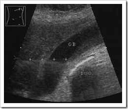
radiology-information.blogspot.com
artifact ultrasound gallbladder artifacts lobe side slice thickness lobes gb imaging physics folio
MRI Artifacts – Susceptibility Artifact – MR-TIP.com
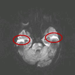
www.mr-tip.com
artifact susceptibility artifacts diffusion mri imaging tip mr visible commonly study case
Digital Radiography Image Artifacts | Radiology | SUNY Upstate Medical
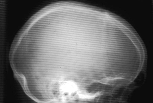
www.upstate.edu
artifact radiography grid radiology digital artifacts aliasing lines cr skull detector lateral upstate medical emaze university burns superimposed barry courtesy
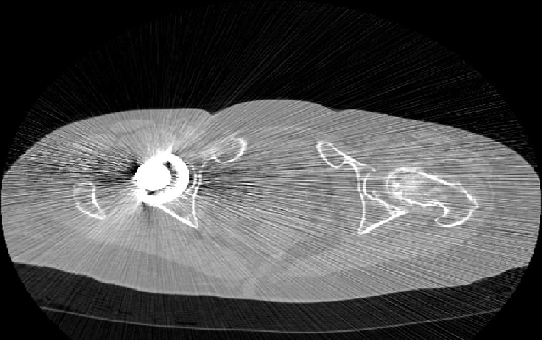
CT Brain With Severe Motion Artifact | Image | Radiopaedia.org

radiopaedia.org
artifact motion brain ct severe movement artifacts radiopaedia patient version
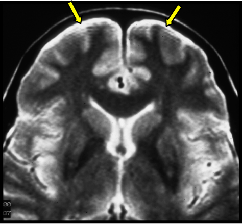
Aliasing Artifact | Image | Radiopaedia.org

radiopaedia.org
artifact aliasing mri artifacts imaging radiology wrap medical radiopaedia caq articles artefacts sequence version
Medical Apparatus Imaging Protocols: MRI

medapparatus.com
mri susceptibility artifact 21a figure protocols references medapparatus
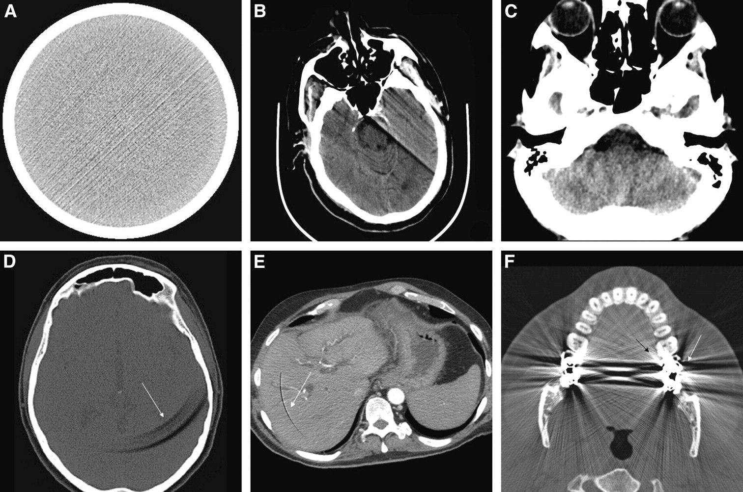
CT Artifacts: Causes And Reduction Techniques
www.openaccessjournals.com
ct artifact ring artifacts imaging medicine techniques reduction head causes figure
Imaging Artifacts | Radiology Key
radiologykey.com
artifacts imaging
MRI Artifacts – Artifact By Patient Movement – MR-TIP.com
www.mr-tip.com
artifact artifacts patient movement mri tip mr causes diffusion imaging study case during
[PDF] Artefacts Found In Computed Radiography. | Semantic Scholar
![[PDF] Artefacts found in computed radiography. | Semantic Scholar](https://d3i71xaburhd42.cloudfront.net/57102cd391e6b9d68d731478ddc620e11471b621/3-Figure3-1.png)
www.semanticscholar.org
computed radiography artefacts
Ultrasound Imaging Guide – Advanced Software Optimization
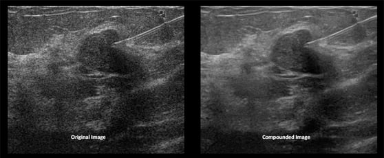
info.umiultrasound.com
ultrasound imaging compounding needle optimization advanced software guide compounded benefits using guidance
Artifacts In Ultrasound Imaging – ECG & ECHO

ecgwaves.com
ultrasound artifacts reverberation artifact imaging sound reflected waves echo boundary illustrated echogenicity layers multiple between times then figure
The Linear Artifact In Enhanced Depth Imaging Spectral Domain Optical
www.nature.com
linear artifact enhanced optical tomography depth spectral coherence imaging domain figure
MRI Artifacts – Ghosting Artifact – MR-TIP.com
www.mr-tip.com
artifact ghosting mri phase artifacts motion tip mr breathing repeating stripes occurring appears result study case
(PDF) Artifact-free High Dynamic Range Imaging
www.researchgate.net
artifact imaging range dynamic
Digital Radiography Image Artifacts | Radiology | SUNY Upstate
www.upstate.edu
radiography radiology artifact artifacts digital cr lines linear reader bright guide light upstate foreign produces output materials figure collection rsna
Imaging Artifacts | Radiology Key

radiologykey.com
artifacts imaging figure
ABC Radiology Blog: Computed Tomography Artifacts
abcradiology.blogspot.com
artifacts streak artifact tomography computed implant radiology
MRI Artifacts – Aliasing Artifact – MR-TIP.com
www.mr-tip.com
aliasing artifact artifacts mri tip mr study case
MRI Artifacts – Eddy Current Artifact – MR-TIP.com
www.mr-tip.com
eddy artifact current artifacts mri tip mr case slice distortion visible whole imaging study over
Dielectric Effect – Questions And Answers In MRI
mri-q.com
dielectric mri artifact effect darkened ascites abdomen 0t abnormally center
Grid Artifact – Jack Frost

jackfrostnowahistoria.blogspot.com
artifact artifacts radiology artefact radiography radiograph radiopaedia upside noted reorder drag
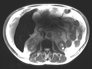
Magnetic Resonance Imaging
www.slideshare.net
resonance
Applicability And Advantages Of Flow Artifact–insensitive Fluid

www.ajnr.org
artifact flow posterior sequences inversion advantages applicability insensitive attenuated fossa recovery fluid imaging mr ajnr
Wraparound Artifact. Axial T2-weighted Single-shot FSE Images (618/180

www.researchgate.net
artifact axial fse t2 weighted wraparound
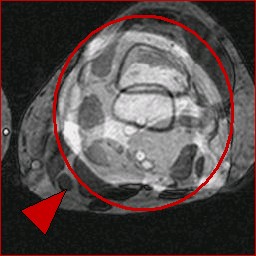
Figure 1: Comparison Of Implant Artifact In Cadaveric Model; Left

www.spinalsurgerynews.com
implants blackarmor artifact spinal surgery tumour engineered artifacts
Digital Radiography Image Artifacts | Radiology | SUNY Upstate
www.upstate.edu
radiography artifacts artifact radiology digital ray collimator upstate medical readout index figure
CAT Scan Image Artifacts
www.ctchiller.com
artifacts artifact scan hardening streak appears blurred
Noise Artifacts In Diagnostic Ultrasound – Innovatus Imaging
www.innovatusimaging.com
ultrasound artifact artifacts
CT Artifacts | Radiology Reference Article | Radiopaedia.org

radiopaedia.org
artifact artifacts radiopaedia radiology cupping radiologija
Artifacts In Ultrasound Imaging – ECG & ECHO

ecgwaves.com
ultrasound artifacts shadowing acoustic imaging artifact shadow echo echocardiography gallstone dark
Aliasing artifact. Artifacts streak artifact tomography computed implant radiology. Cat scan image artifacts